What Baby Looks Like in Utero at 38 Weeks
| Obstetric ultrasonography | |
|---|---|
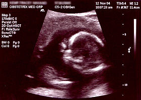 Obstetric sonogram of a fetus at xvi weeks. The bright white circumvolve center-correct is the head, which faces to the left. Features include the brow at x o'clock, the left ear toward the center at 7 o'clock and the right manus covering the optics at nine:00. | |
| Other names | prenatal ultrasound |
| ICD-9-CM | 88.78 |
| MeSH | D016216 |
| OPS-301 code | iii-032, 3-05d |
Obstetric ultrasonography, or prenatal ultrasound, is the employ of medical ultrasonography in pregnancy, in which audio waves are used to create real-time visual images of the developing embryo or fetus in the uterus (womb). The procedure is a standard part of prenatal intendance in many countries, as it can provide a variety of information most the health of the mother, the timing and progress of the pregnancy, and the health and evolution of the embryo or fetus.
The International Order of Ultrasound in Obstetrics and Gynecology (ISUOG) recommends that pregnant women have routine obstetric ultrasounds betwixt 18 weeks' and 22 weeks' gestational age (the anatomy scan) in order to confirm pregnancy dating, to measure out the fetus so that growth abnormalities can be recognized speedily later in pregnancy, and to appraise for built malformations and multiple pregnancies (twins, etc).[1] Additionally, the ISUOG recommends that pregnant patients who desire genetic testing have obstetric ultrasounds between 11 weeks' and 13 weeks 6 days' gestational age in countries with resource to perform them (the nuchal scan). Performing an ultrasound at this early on stage of pregnancy can more accurately confirm the timing of the pregnancy, and can likewise assess for multiple fetuses and major built abnormalities at an earlier stage.[2] Research shows that routine obstetric ultrasound before 24 weeks' gestational age tin significantly reduce the take a chance of declining to recognize multiple gestations and can meliorate pregnancy dating to reduce the risk of labor induction for post-dates pregnancy. There is no deviation, however, in perinatal death or poor outcomes for infants.[3]
Terminology [edit]

Below are useful terms on ultrasound:[four]
- Echogenic — giving rise to reflections (echoes) of ultrasound waves
- Hyperechoic – more echogenic (brighter) than normal
- Hypoechoic – less echogenic (darker) than normal
- Isoechoic – the aforementioned echogenicity every bit another tissue
- Transvaginal ultrasonography – Ultrasound is performed through the vagina
- Transabdominal ultrasonography – Ultrasound is performed beyond the abdominal wall or through the intestinal cavity
In normal state, each torso tissue type, such equally liver, spleen or kidney, has a unique echogenicity. Fortunately, gestational sac, yolk sac and embryo are surrounded by hyperechoic (brighter) body tissues.
Types [edit]
Traditional obstetric sonograms are done by placing a transducer on the abdomen of the meaning woman. One variant, transvaginal sonography, is done with a probe placed in the woman's vagina. Transvaginal scans unremarkably provide clearer pictures during early pregnancy and in obese women. As well used is Doppler sonography which detects the heartbeat of the fetus. Doppler sonography can be used to evaluate the pulsations in the fetal heart and bloods vessels for signs of abnormalities.[5]
3D ultrasound [edit]
Modernistic 3D ultrasound images provide greater item for prenatal diagnosis than the older 2D ultrasound technology.[6] While 3D is popular with parents desiring a prenatal photograph as a keepsake,[7] both 2D and 3D are discouraged by the FDA for non-medical use,[8] but there are no definitive studies linking ultrasound to any adverse medical furnishings.[9] The post-obit 3D ultrasound images were taken at unlike stages of pregnancy:
-
3D Ultrasound of fetal movements at 12 weeks
-

Fetus at 17 weeks
-

Fetus at xx weeks
Medical uses [edit]
Early on pregnancy [edit]
A gestational sac tin can be reliably seen on transvaginal ultrasound by five weeks' gestational age (approximately 3 weeks later ovulation). The embryo should be seen past the time the gestational sac measures 25 mm, most five-and-a-one-half weeks.[10] The heartbeat is usually seen on transvaginal ultrasound by the time the embryo measures 5 mm, but may non be visible until the embryo reaches 19 mm, around 7 weeks' gestational age.[v] [eleven] [12] Coincidentally, near miscarriages also happen by 7 weeks' gestation. The rate of miscarriage, especially threatened miscarriage, drops significantly later normal heartbeat is detected, and afterward thirteen weeks.[xiii]
-
Embryo at 5 weeks and i day of gestational historic period (at top left) with discernible heartbeat.
-
Embryo at 5 weeks and 5 days of gestational historic period with discernible heartbeat.
First trimester [edit]
In the showtime trimester, a standard ultrasound examination typically includes:[12]
- Gestational sac size, location, and number
- Identification of the embryo and/or yolk sac
- Measurement of fetal length (known as the crown-rump length)
- Fetal number, including number of amnionic sacs and chorionic sacs for multiple gestations
- Embryonic/fetal cardiac activeness
- Assessment of embryonic/fetal anatomy appropriate for the beginning trimester
- Evaluation of the maternal uterus, tubes, ovaries, and surrounding structures
- Evaluation of the fetal nuchal fold, with consideration of fetal nuchal translucency assessment
Second and third trimester [edit]
In the 2d trimester, a standard ultrasound exam typically includes:[12]
- Fetal number, including number of amnionic sacs and chorionic sacs for multiple gestations
- Fetal cardiac action
- Fetal position relative to the uterus and cervix
- Location and appearance of the placenta, including site of umbilical cord insertion when possible
- Amnionic fluid volume
- Gestational age assessment
- Fetal weight estimation
- Fetal anatomical survey
- Evaluation of the maternal uterus, tubes, ovaries, and surrounding structures when advisable
Dating and growth monitoring [edit]
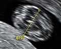
Biparietal bore is taken as the maximal transverse bore of in a visualization of the horizontal airplane of the caput.

Gestational age is usually determined past the date of the adult female's last menstrual menstruation, and bold ovulation occurred on mean solar day 14 of the menstrual bicycle. Sometimes a woman may exist uncertain of the date of her last menstrual period, or there may be reason to suspect ovulation occurred significantly earlier or later than the fourteenth 24-hour interval of her bike. Ultrasound scans offer an culling method of estimating gestational age. The near accurate measurement for dating is the crown-rump length of the fetus, which can be done between 7 and 13 weeks of gestation. Later on thirteen weeks of gestation, the fetal historic period may be estimated using the biparietal diameter (the transverse diameter of the caput, across the two parietal basic), the head circumference, the length of the femur, the crown-heel length (head to heel), and other fetal parameters.[ commendation needed ] Dating is more accurate when washed before in the pregnancy; if a later scan gives a different gauge of gestational age, the estimated age is not normally changed but rather it is assumed the fetus is not growing at the expected rate.[five]
The intestinal circumference of the fetus may also be measured. This gives an estimate of the weight and size of the fetus and is important when doing series ultrasounds to monitor fetal growth.[5]
Fetal sex discernment [edit]
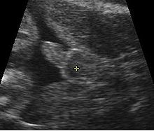
Sonogram of male fetus, with scrotum and penis in heart of image
The sexual practice of the fetus may be discerned by ultrasound equally early equally 11 weeks' gestation. The accuracy is relatively imprecise when attempted early.[15] [16] [17] Afterward 13 weeks' gestation, a high accuracy of between 99% and 100% is possible if the fetus does not brandish intersex external characteristics.[eighteen]
The post-obit is accurateness data from two hospitals:
| Gestational Age | Male monarch's College Hospital Medical Schoolhouse[16] | Taipei City Hospital & Li Shin Hospital[17] |
|---|---|---|
| eleven weeks | 70.iii% | 71.9% |
| 12 weeks | 98.7% | 92% |
| 13 weeks | 100% | 98.iii% |
Influencing factors [edit]
The accurateness of fetal sexual practice discernment depends on:[15]
- Gestational age
- Precision of sonographic machine
- Expertise of the operator
- Fetal posture
Ultrasonography of the cervix [edit]
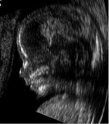
Fetus at 14 weeks (contour)

Fetus at 14 weeks with avant-garde imaging filters
Obstetric sonography is useful in the assessment of the cervix in women at risk for premature birth. A brusk neck preterm is associated with a higher take a chance for premature delivery: At 24 weeks' gestation, a cervix length of less than 25 mm defines a risk group for spontaneous preterm birth. Further, the shorter the cervix, the greater the take a chance.[19] Cervical measurement on ultrasound too has been helpful to use ultrasonography in patients with preterm contractions, as those whose cervical length exceeds 30 mm are unlikely to deliver within the next week.[xx]
Abnormality screening [edit]
In near countries, routine pregnancy sonographic scans are performed to detect developmental defects before birth. This includes checking the status of the limbs and vital organs, likewise as (sometimes) specific tests for abnormalities. Some abnormalities detected by ultrasound can be addressed by medical treatment in utero or past perinatal care, though indications of other abnormalities tin can lead to a determination regarding abortion.
Maybe the most common such test uses a measurement of the nuchal translucency thickness ("NT-test", or "Nuchal Browse"). Although 91% of fetuses affected by Downward syndrome exhibit this defect, 5% of fetuses flagged past the test do non have Down syndrome.
Ultrasound may also observe fetal organ anomaly. Ordinarily scans for this type of detection are washed around xviii to 23 weeks of gestational age (called the "anatomy scan", "anomaly browse," or "level 2 ultrasound"). Some resource signal that there are clear reasons for this and that such scans are also conspicuously beneficial considering ultrasound enables clear clinical advantages for assessing the developing fetus in terms of morphology, bone shape, skeletal features, fetal heart office, volume evaluation, fetal lung maturity,[21] and general fetus well beingness.[22]
Second-trimester ultrasound screening for aneuploidies is based on looking for soft markers and some predefined structural abnormalities. Soft markers are variations from normal beefcake, which are more common in aneuploid fetuses compared to euploid ones. These markers are oft non clinically significant and practise non cause adverse pregnancy outcomes.[23]
Safe issues [edit]
3D rendering of the fetal spine in a scan at nineteen weeks of pregnancy
Current evidence indicates that diagnostic ultrasound is safe for the unborn kid, dissimilar radiographs, which employ ionizing radiation. Randomized controlled trials accept followed children up to ages eight–9, with no pregnant differences in vision, hearing, school functioning, dyslexia, or spoken language and neurologic development by exposure to ultrasound.[24] In one randomized trial, the children with greater exposure to ultrasound had a reduction in perinatal mortality, and was attributed to the increased detection of anomalies in the ultrasound group.[24]
The 1985 maximum power immune by the U.South. Food and Drug Administration (FDA) of 180 milliwatts per square cm[25] is well nether the levels used in therapeutic ultrasound, but notwithstanding college than the 30-fourscore milliwatts per square cm range of the Statison 5 veterinary LIPUS device.[26]
Doppler ultrasonography examinations has a thermal index (TI) of about five times that of regular (B-mode) ultrasound examinations.[24] Several randomized controlled trials accept reported no association between Doppler exposure and birth weight, Apgar scores, and perinatal mortality. One randomized controlled trial, however, came to the result of a higher perinatal death rate of usually formed infants born after 24 weeks exposed to Doppler ultrasonography (RR 3.95, 95% CI i.32–xi.77), but this was non a primary effect of the written report, and has been speculated to be due to take chances rather than a harmful issue of Doppler itself.[24]
The FDA discourages its use for non-medical purposes such as fetal keepsake videos and photos, even though it is the same technology used in hospitals.[27]
The American Institute of Ultrasound in Medicine recommends spectral Doppler only if M-mode sonography is unsuccessful, and even then simply briefly, due to the acoustic intensity delivered to the fetus.[28]
History [edit]
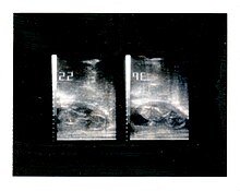
Polaroid photo of an obstetric ultrasound taken in 1985.
Scottish physician Ian Donald was one of the pioneers of medical use of ultrasound. His article "Investigation of Intestinal Masses by Pulsed Ultrasound" was published in The Lancet in 1958.[29] Donald was Regius Professor of Midwifery at the University of Glasgow.[thirty] [ self-published source? ]
In 1962, David Robinson, George Kossoff, George Radovanovich, and Dr William Garrett were the start in the world to identify a number of foetal anatomical structures from high frequency sound wave imaging.[31] [32]
In 1962, after near ii years of piece of work, Joseph Holmes, William Wright, and Ralph Meyerdirk developed the outset chemical compound contact B-mode scanner. Their work had been supported past U.S. Public Health Services and the University of Colorado. Wright and Meyerdirk left the academy to form Physionic Engineering Inc., which launched the first commercial hand-held articulated arm compound contact B-mode scanner in 1963.[33] [ cocky-published source? ] This was the starting time of the most popular design in the history of ultrasound scanners.
Obstetric ultrasound has played a significant function in the evolution of diagnostic ultrasound engineering science in general. Much of the technological advances in diagnostic ultrasound technology are due to the drive to create better obstetric ultrasound equipment. Acuson Corporation's pioneering piece of work on the development of Coherent Image Formation helped shape the development of diagnostic ultrasound equipment as a whole.[ commendation needed ]
In March and April 2015, a mail by a meaning woman named Jen Martin (née Cardinal) and her husband to YouTube, which had been viewed at least ii million times and had many likes, showed the 14-week-old fetus clapping repeatedly to the song, sung past the parents, "If You're Happy And Yous Know Information technology." Information technology was later revealed that the video- while not a false- had been somewhat edited to testify more than fetal claps than likely occurred. It is not unprecedented for fetuses of that historic period to make momentary movements that could be repeated once or twice beyond the initial movement, according to experts, but to echo such a move more than that- especially purposefully- would non likely be feasible at that point.[34] [35] [36]
Society and culture [edit]
The increasingly widespread use of ultrasound technology in monitoring pregnancy has had a neat impact on the way in which women and societies at big conceptualise and feel pregnancy and childbirth.[37] The pervasive spread of obstetric ultrasound technology around the world and the conflation of its use with creating a 'safe' pregnancy besides as the ability to meet and determine features like the sex of the fetus bear on the way in which pregnancy is experienced and conceptualised.[37] This "technocratic takeover"[37] of pregnancy is non express to western or developed nations only also affects conceptualisations and experiences in developing nations and is an case of the increasing medicalisation of pregnancy, a miracle that has social as well as technological ramifications.[37] Ethnographic enquiry concerned with the use of ultrasound technology in monitoring pregnancy tin can bear witness us how it has changed the embodied experience of expecting mothers around the globe.[37]
Recent studies take stressed the importance of framing "reproductive health matters cross-culturally", particularly when agreement the "new miracle" of "the proliferation of ultrasound imaging" in developing countries.[38] In 2004, Tine Gammeltoft interviewed 400 women in Hanoi'due south Obstetrics and Gynecology Hospital; each "had an average of six.6 scans during her pregnancy", much higher than v years prior when "a pregnant adult female might or might not have had a single scan during her pregnancy" in Vietnam.[38] Gammeltoft explains that "many Asian countries" see "the foetus as an ambiguous being" unlike in Western medicine where it is common to think of the foetus equally "materially stable".[38] Therefore, although women, particularly in Asian countries, "express intense uncertainties regarding the safety and credibility of this engineering science", it is overused for its "firsthand reassurance".[38]
Run into also [edit]
- 3D ultrasound
- Doppler fetal monitor
- Global Library of Women's Medicine
- Gynecologic ultrasonography
References [edit]
- ^ Salomon, LJ; Alfirevic, Z; Berghella, 5; Bilardo, C; Hernandez-Andrade, E; Johnsen, SL; Kalache, K; Leung, K.-Y.; Malinger, G; Munoz, H; Prefumo, F; Toi, A; Lee, Due west (2010). "Practice guidelines for performance of the routine mid-trimester fetal ultrasound scan". Ultrasound Obstet Gynecol. 37 (one): 116–126. doi:10.1002/uog.8831. PMID 20842655. S2CID 10676445.
- ^ Salomon, LJ; Alfirevic, Z; Bilardo, CM; Chalouhi, GE; Ghi, T; Kagan, KO; Lau, TK; Papageorghiou, AT; Raine-Fenning, NJ; Stirnemann, J; Suresh, S; Tabor, A; Timor-Tritsch, IE; Toi, A; Yeo, G (2013). "ISUOG Practice Guidelines: operation of first-trimester fetal ultrasound scan". Ultrasound Obstet Gynecol. 41 (i): 102–113. doi:10.1002/uog.12342. PMID 23280739. S2CID 13593.
- ^ Whitworth, M; Bricker, L; Mullan, C (2015). "Ultrasound for fetal assessment in early pregnancy". Cochrane Database of Systematic Reviews (7): CD007058. doi:10.1002/14651858.CD007058.pub3. PMC4084925. PMID 26171896.
- ^ Zwingenberger, Allison (10 April 2007). "What do hyperechoic and hypoechoic mean?". DVM Journals.
- ^ a b c d Woo, Joseph (2006). "Why and when is Ultrasound used in Pregnancy?". Obstetric Ultrasound: A Comprehensive Guide . Retrieved 2007-05-27 .
- ^ Dimitrova V, Markov D, Dimitrov R (2007). "[3D and 4D ultrasonography in obstetrics]". Akush Ginekol (Sofiia) (in Bulgarian). 46 (2): 31–twoscore. PMID 17469450.
- ^ Sheiner Eastward, Hackmon R, Shoham-Vardi I, et al. (2007). "A comparison between acoustic output indices in 2D and 3D/4D ultrasound in obstetrics". Ultrasound Obstet Gynecol. 29 (3): 326–viii. doi:10.1002/uog.3933. PMID 17265534. S2CID 41853089.
- ^ Rados C (January–February 2004). "FDA Cautions Against Ultrasound 'Keepsake' Images". FDA Consumer Magazine. Archived from the original on xiii May 2009. Retrieved 28 February 2012.
- ^ Kempley R (9 August 2003). "The Grin Before They Bear It; Peek-a-Boo: Prenatal Portraits for the Ultrasound Set". Washington Post. Archived from the original on ii November 2012.
- ^ Doubilet, Peter M.; Benson, Carol B.; Bourne, Tom; Blaivas, Michael (2013-10-ten). Campion, Edward Westward. (ed.). "Diagnostic Criteria for Nonviable Pregnancy Early in the First Trimester". New England Journal of Medicine. 369 (xv): 1443–1451. doi:10.1056/NEJMra1302417. ISSN 0028-4793. PMID 24106937.
- ^ Boschert, Sherry (2001-06-xv). "Anxious Patients Oftentimes Want Very Early Ultrasound Exam". OB/GYN News. FindArticles.com. Retrieved 2007-05-27 .
- ^ a b c Cunningham, F; Leveno, KJ; Bloom, SL; Spong, CY; Dashe, JS; Hoffman, BL; Casey BM, BM; Sheffield, JS (2013). "Fetal Imaging". Williams Obstetrics, 20-Quaternary Edition. McGraw-Hill.
- ^ "Miscarriage". A.D.A.One thousand., Inc. 21 November 2010. Retrieved 28 February 2012.
- ^ Snijders, RJ.; Nicolaides, KH. (Jan 1994). "Fetal biometry at fourteen-40 weeks' gestation". Ultrasound Obstet Gynecol. 4 (1): 34–48. doi:10.1046/j.1469-0705.1994.04010034.ten. PMID 12797224. S2CID 19399509.
- ^ a b Merz, Eberhard (2005). Ultrasound in obstetrics and gynecology (2nd ed.). Stuttgart: Thieme. p. 129. ISBN978-1-58890-147-7.
- ^ a b Efrat, Z.; Akinfenwa, O. O.; Nicolaides, K. H. (1999). "First-trimester determination of fetal gender past ultrasound". Ultrasound in Obstetrics and Gynecology. 13 (5): 305–vii. doi:ten.1046/j.1469-0705.1999.13050305.10. PMID 10380292. S2CID 5364077.
- ^ a b Hsiao, C.H.; Wang, H.C.; Hsieh, C.F.; Hsu, J.J. (2008). "Fetal gender screening by ultrasound at 11 to 13+half-dozen weeks". Acta Obstetricia et Gynecologica Scandinavica. 87 (ane): 8–13. doi:10.1080/00016340701571905. PMID 17851807. S2CID 22374986.
- ^ Odeh, Marwan; Grinin, Vitali; Kais, Mohamad; Ophir, Ella; Bornstein, Jacob (2009). "Sonographic Fetal Sexual practice Determination". Obstetrical & Gynecological Survey. 64 (1): 50–57. doi:10.1097/OGX.0b013e318193299b. PMID 19099612. S2CID 205898633.
- ^ Iams, Jay D.; Goldenberg, Robert L.; Meis, Paul J.; Mercer, Brian Grand.; Moawad, Atef; Das, Anita; Thom, Elizabeth; McNellis, Donald; et al. (1996). "The Length of the Cervix and the Risk of spontaneous Premature Delivery". New England Journal of Medicine. 334 (9): 567–72. doi:10.1056/NEJM199602293340904. PMID 8569824.
- ^ Leitich, Harald; Brunbauer, Mathias; Kaider, Alexandra; Egarter, Christian; Husslein, Peter (1999). "Cervical length and dilatation of the internal cervical os detected by vaginal ultrasonography as markers for preterm delivery: A systematic review". American Journal of Obstetrics and Gynecology. 181 (6): 1465–72. doi:10.1016/S0002-9378(99)70407-2. PMID 10601930.
- ^ Bhanu Prakash, 1000.N.; Ramakrishnan, A.G.; Suresh, South.; Chow, T.W.P. (March 2002). "Fetal lung maturity analysis using ultrasound image features" (PDF). IEEE Transactions on Information technology in Biomedicine. 6 (1): 38–45. doi:10.1109/4233.992160. PMID 11936595. S2CID 14662967.
- ^ Layyous, Najeeb. "The Clinical Advantages of 3D and 4D Ultrasound - Dr Due north Layyous". www.layyous.com . Retrieved 21 March 2018.
- ^ Zare Mehrjardi, Mohammad; Keshavarz, Elham (2017-04-16). "Prefrontal Space Ratio—A Novel Ultrasound Marking in the 2nd Trimester Screening for Trisomy 21: Systematic Review and Meta-Assay". Journal of Diagnostic Medical Sonography. 33 (four): 269–277. doi:10.1177/8756479317702619.
- ^ a b c d Houston, Laura E.; Odibo, Anthony O.; Macones, George A. (2009). "The rubber of obstetrical ultrasound: a review". Prenatal Diagnosis. 29 (thirteen): 1204–1212. doi:ten.1002/pd.2392. ISSN 0197-3851. PMID 19899071. S2CID 26980283.
- ^ Freitas, Robert A. (1999). Nanomedicine. Austin, TX: Landes Bioscience. ISBN978-1-57059-645-ii. [ folio needed ]
- ^ "Statison 5 Operations Manual" (PDF). Statison Medical, Inc. 1997. Archived from the original (PDF) on 27 May 2008.
- ^ "Fetal Emblem Videos". Nutrient and Drug Administration. Retrieved 2011-05-21 .
- ^ "Argument on Measurement of the Fetal Centre Charge per unit". Sound Waves Weekly. American Institute of Ultrasound in Medicine. November 17, 2011.
When attempting to obtain fetal heart charge per unit with a diagnostic ultrasound system, the AIUM recommends using Thou-mode at get-go considering the time-averaged audio-visual intensity delivered to the fetus is lower with M-mode than with spectral Doppler. If this is unsuccessful, spectral Doppler ultrasound may be used with the following guidelines: use spectral Doppler only briefly (eg, 4-five heart beats), and keep the thermal index (TIS for soft tissues in the beginning trimester and TIB for bones in second and tertiary trimesters) as low as possible, preferably beneath 1 in accordance with the ALARA (as low as reasonably achievable) principle.
- ^ Donald, I; MacVicar, J; Chocolate-brown, TG (1958). "Investigation of abdominal masses by pulsed ultrasound". Lancet. one (7032): 1188–95. doi:10.1016/S0140-6736(58)91905-6. PMID 13550965.
- ^ Ian Donald'due south paper in the Lancet in 1958 past Joseph Woo
- ^ "History of Sonography in Commonwealth of australia". Retrieved 17 Baronial 2018.
- ^ "Bill Garrett: Obstetrics practitioner helped develop ultrasound". The Sydney Morning Herald. 10 December 2015. Retrieved 17 August 2018.
- ^ Woo, Joseph (2002). "A brusque History of the development of Ultrasound in Obstetrics and Gynecology". ob-ultrasound.net. Retrieved 2007-08-26 .
- ^ "Ultrasound Appears To Show Fetus Clapping To 'If You're Happy And You Know It'". Huffington Mail service. 30 March 2015.
- ^ "Ultrasound Shows Baby Clapping To 'If You're Happy And Yous Know It'". inquisitr.com. 28 March 2015. Retrieved 21 March 2018.
- ^ "Archived copy". Archived from the original on 2015-04-xiii. Retrieved 2015-04-04 .
{{cite web}}: CS1 maint: archived copy as title (link) - ^ a b c d east [Gammeltoft, Tine, 2007, Sonography and Sociality – Obstetrical Ultrasound Imagining in Urban Vietnam, Medical Anthropology Quarterly, 21:two, 133-153]
- ^ a b c d Gammeltoft, Tine (2007). "Sonography and Sociality: Obstetrical Ultrasound Imaging in Urban Vietnam". Medical Anthropology Quarterly. 21 (2): 133–53. doi:10.1525/maq.2007.21.2.133. PMID 17601081.
External links [edit]
- RadiologyInfo: Obstetric Ultrasound Imaging
- AIUM argument on prudent utilize of Ultrasound
- The Global Library of Women's Medicine Imaging in Obstetrics and Gynecology link
Source: https://en.wikipedia.org/wiki/Obstetric_ultrasonography
0 Response to "What Baby Looks Like in Utero at 38 Weeks"
Post a Comment